vol. 6 2/2017 Inżynier i Fizyk Medyczny
90
radiologia
\
radiology
artykuł naukowy
\
scientific paper
AEC System Testing
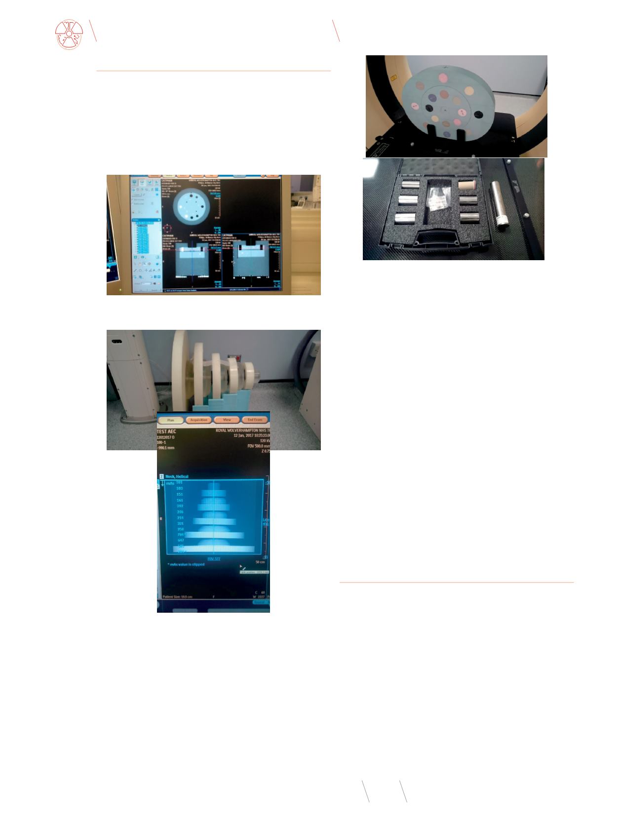
The AEC system has been tested using phantoms: CATPHAN 600
(image quality - SD, SNR, CNR, contrast, uniformity and quantity
- HU) (Fig.2.), Philips test object built from 5 circle phantoms with
separations: 15cm, 20cm, 26.5cm, 36.5cm, 50cm (image quality and
quantity) (Fig.3), CIRS with additional inserts of high density mate-
rials: aluminum, titanium, stainless steel (image quantity) (Fig. 4.).
Fig. 3
The Philips test object and the scannogram
Source: Authors’ materials.
Fig. 2
The CT scan of the CATPHAN600
Source: Authors’ materials.
Fig. 4
CIRS phantom and high density inserts – aluminum, titanium, stainless steel
Source: Authors’ materials.
The CIRS phantomwas used to verify the AEC system in terms
of HU values for particular materials (image quantity).
Testing conditions were: 120kV for all protocols, 2/3mm slice
width, 2/3mm reconstruction increment, the scanner standard re-
construction kernel, reconstruction FOV (RFOV) dictated by setting
of clinical protocols (50-70 cm), B/UB filters (brain protocol/rest of
body protocols), all DoseRight facilities ON, iDose reconstruction
algorithm at a level 3. All used phantoms were carefully aligned
parallel to the scan plane and centered in the field of view (FOV).
The systemwas tested for helical protocols as that kindof protocols
will be used in a clinical practice. All tested protocols were tested
for fixed mAs as well as the AEC switched ON. A scan series was se-
lected that covered the length and separation of the phantoms. For
patient sizes AEC, a range of patient sizes were simulated and as-
sessed using Surview functionality (A-P and LAT scannogram). The
system “mapped” mAs along z-axis in those projections.
The effect of changing the AEC image quality level and quantity
parameters were assessed in relation to fixed protocols and among
them. There was also assessed a long term stability of the system.
Image quality and quantity assessment
The images from the testing were assessed. The CATPHAN600
images for fixed mAs (tab.1.) and ACS (tab.2.) protocols were an-
alysed using the image quality parameters: uniformity, contrast,
CNR, SD and image quantity: HU.
Uniformity for both types of protocols – fixed mAs and ACS
– for particular anatomical regions is similar and doesn’t show
any trends in terms of exposure, protocols setups, scanning and
reconstruction facilities. It can depend on what the reference
image is chosen and determined by a setup for each protocol.
Contrast and CNR for the fixed protocols is better but it can be
resulted by higher exposure in an average way and lack of depen-
dency on the reference image parameters. It is also an effect of
a compromise between image quality and provided dose. The HU
The circle shaped AEC Philips phantom is manufactured from
plastic material, which has no significantly lower density than
water (-35 HU). The object contains 5 circle uniform objects and
increases in area along the z-axis. Originally that object was
used to calibrate older models of Philps/Elscint CT scanners as
well as Philips standard simulators. The object was used to test
the AEC system to prove its appropriate working in relation to
the patient size and z-axis. A design of that object causes a high
modulation of the mA of the x-ray tube when during scanning.


