Inżynier i Fizyk Medyczny 6/2016 vol. 5
319
artykuł
/
article
radioterapia
/
radiotherapy
. A brachytherapy applicator (Rotherham system – ovoids, ring
applicator, VSA) is placed in the uterus/vagina during a theatre
procedure. The images from these two modalities are fused to
provide needed image information to mark up the treated volu-
mes (GTV, HRV, IRV) and OARs (rectum, sigmoid, bowels, blad-
der). 3D planning additionally confirms of applicator placement,
gives possibilities to decrease OARs dose for patients with
a small cervix, accounting for sigmoid colon dose, and improves
coverage for large volume disease (wide infiltration of disease
into a tissue) while maintaining OARs’ dosimetry. At the time
of brachytherapy application, tandem placement can result in
unsuspected uterine perforation despite the clinical impression
of adequate tandem placement. Additionally, introducing MRI
has given optimization to maintain tumour coverage and reduce
the dose to the normal tissues, especially in patients with small
cervix where the target volume treated.
The EMBRACE, RCR and GEC-ESTRO guides recommenda-
tions are used on contouring of tumour targets and OARs as
well as dose volume parameters to be reported for IGBT (Image
Guided Brachytherapy) for locally advanced cervix cancer. The
dose reported to the target volume (90% of volume GTV, HRV,
IRV – D90)as well as the OARs’ volumes (0.1cc, 1.0cc, 2.0cc of
OAR volume) is calculated using EQD2 to combine the ERT dose
and BT dose. EQD2 is also reported to A-points to collect eviden-
ces to compare an “old” techniques prescription (dose norma-
lisation to A-points) and a volumetric optimisation technique.
The optimisation dose distribution is very difficult in terms of
getting the recommended doses prescriptions with keeping
constraints for OARs.
When the treatment plan is accepted by a consultant and
checked independently by a second physicist, the patient is pla-
ced in the treatment room and the applicator is connected via
a transfer tube to the Flexitron treatment delivery unit (TDU).
Radiation dose is delivered via a single source (Ir-192) welded
at the end of a steel cable, driven to the number of planned
dwell positions and dwell times within the applicator for a pe-
riod about 10-15 minutes creating desired dose distribution.
The source then moves into the TDU safe for about 1 hour. The
planned number of repetitions is 26-35 pulses and prescribed
dose per pulse is 0.8-1.0 Gy. The patient remains a bed bound
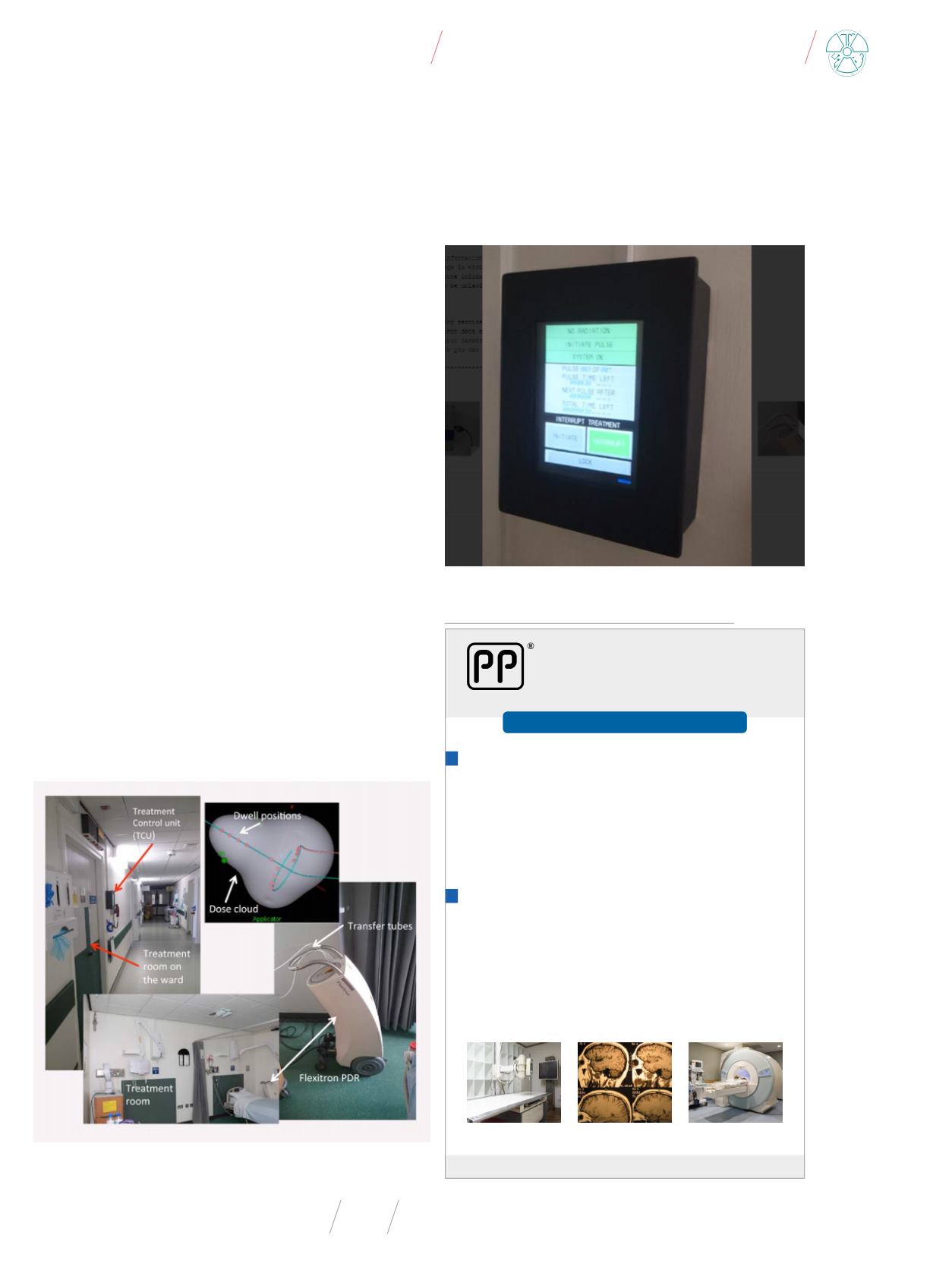
Fig. 2
. The PDR treatment system and treatment room
Source: [4] and own materials.
Fig. 3
. The control unit of the PDR system
Source: Own materials.
ISTNIEJEOD1989R.
OŚRODEK BADAŃ i ANALIZ „PP”
Marek Zając i Artur Zając s.c.
ul. prof. Michała Bobrzyńskiego 23A/U2, 30-348 KRAKÓW,
fax: +48 12 202 04 77, tel.: +48 603 18 77 88,
e-mail:
– testy specjalistyczne aparatury rentgenowskiej (stomatologia,
radiografia, fluoroskopia, mammografia, tomografia komputerowa),
– pomiary dozymetryczne w środowisku pracy i w środowisku
w otoczeniu aparatów rtg,
– projekty pracowni rtg wraz z obliczaniem osłon stałych,
– szkolenia z zakresu wykonywania testów podstawowych,
– opracowujemy dokumentację Systemu Jakości w pracowniach rtg.
– natężenia pola elektromagnetycznego (m.in. rezonans magnetyczny),
– hałasu i drgań
– natężenia i równomierności oświetlenia na stanowiskach pracy i oświetlenia
awaryjnego,
– promieniowania optycznego nielaserowego (180–3000 nm): nadfioletowe,
widzialne (w tym niebieskie), podczerwone,
– promieniowania laserowego,
– pobieranie prób powietrza oraz oznaczanie zawartości pyłu
całkowitego i respirabilnego.
WYKONUJEMY
PONADTO WYKONUJEMY POMIARY:
POSIADAMY AKREDYTACJĘ NR AB 286
reklama


